| front |1 |2 |3 |4 |5 |6 |7 |8 |9 |10 |11 |12 |13 |14 |15 |16 |17 |18 |19 |20 |21 |22 |23 |24 |review |
 |
Several
age-associated cardiovascular changes directly impact the development and
progression of atherosclerosis and CAD.1 Over a century ago, it
was recognized that arterial walls stiffen with age in both animals and man.
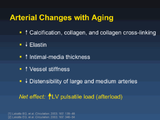
Major aging changes in the arterial wall include increased calcium and
collagen content and loss of elastin fibers. An increase in collagen
cross-linking also occurs. Greater wall thickness is observed in both
central and peripheral arteries and can be detected by ultrasound as
increased intima-medial thickness. The loss in arterial resiliency results
in increases in the systolic blood pressure and pulse pressure, both of
which are potent cardiovascular and coronary risk factors in older patients.
Pulse wave velocity, a more direct measure of arterial stiffness, also
increases with age. The central arterial pressure wave contour is also
altered with age, typically demonstrating late systolic augmentation,
thought to represent the effects of early wave reflection returning from the
periphery. Stiffer arteries with higher systolic and pulse pressures coupled
with late systolic pressure augmentation combine to increase the left
ventricular (LV) pulsatile load with age. Thus, the older adult heart requires greater LV stroke work, wall tension and myocardial oxygen consumption during systole than does the younger heart, thereby increasing the risk of ischemia, even in the absence of CAD. In addition, vascular wall stress is increased, predisposing to vascular injury, an important stimulus to the development and progression of atherosclerosis. [1] Lakatta EG, et al. Arterial and cardiac aging: major shareholders in cardiovascular disease enterprises part I: aging arteries: a set up for vascular disease. Circulation. 2003; 107:139146 [2] Lakatta EG, et al. Arterial and cardiac aging: major shareholders in cardiovascular disease enterprises part II: the aging heart in health: links to heart disease. Circulation. 2003; 107:346354 |